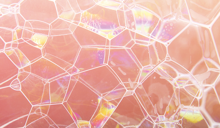
Photograph of scutoid shape

Can the Scutoid Shape the Future of Regenerative Medicine?

The discovery of a new three-dimensional shape could advance understanding of cell topology and the field of regenerative medicine.

Story by

Lori Friedman

Photography by

Christa Neu (main)

Videography by

Stephanie Veto

During development, tissue made of epithelial cells bends to form complex, three-dimensional structures. The cells curve and pack tightly together to accommodate the bending. Researchers have assumed that epithelial cells retain a uniform shape in three dimensions. Through mathematical modeling and experimental research, biophysicist Javier Buceta and an international group of scientists have discovered that, instead, epithelial cells morph into novel shapes that are not the same from top to bottom.

Buceta, an associate professor of bioengineering and also of chemical and biomolecular engineering, and his colleagues dubbed this new three-dimensional (3-D) shape the scutoid.

The identification of the scutoid was named one of the top math stories of 2018 by the American Mathematical Society. Discover Magazine’s State of Science 2019 highlighted the finding as a notable mathematical breakthrough. Scutoids quickly became a pop-culture sensation on Twitter and late-night television as the public humorously fretted for future middle-schoolers who would have to calculate the volume of a scutoid, which Buceta described as “a twisted prism with a zipper.”

photograph of soap bubbles pressed together in scutoid shapes

Buceta and his colleagues named the new shape the scutoid because of its resemblance to a beetle body part called the scutellum. Physicists have discovered scutoids in a bubbly foam.

Yet, in the long run, the discovery may be most notable for its contribution to the field of biomedicine.

The team hypothesized that epithelial cells adopt scutoid shapes in order to pack themselves as efficiently as possible, following a physics principle known as energy minimization. Since then, their hypothesis has been independently validated by the work of an unaffiliated group of researchers that, through a controlled experiment, observed soap bubbles forming scutoid shapes in order to minimize their surface energies.

The identification of epithelial cells’ 3-D structure has wide-reaching implications for advancing the scientific community’s understanding of cellular topology, as well as the development of artificially-grown tissues and organs.

A New Paradigm to Understand Cell Health

“It has been known for a while that a lot can be learned about the health of a cell by analyzing its geometry—its shape and size—and topology—its arrangement with neighboring cells,” explains Buceta. “Healthy cells follow a certain distribution pattern. A change in this distribution pattern can be observed in unhealthy cells.”

An analytical model that assumes a uniform structure is no longer sufficient, says Buceta. The team is interested in delving further into this area in order to develop new tools that characterize tissues using information about epithelial cells’ three-dimensional shape.

“It is now possible to conduct a more realistic analysis that will consider the different cellular relationships and mechanical interactions between the epithelial cells in normal and pathological conditions,” adds Buceta’s collaborator, Luis M. Escudero of the University of Seville in Spain.

“Despite significant progress, tissue engineering and regenerative medicine still falls short when it comes to producing tissues that are duplicates of the native counterpart,” says Buceta’s Lehigh colleague, Lesley Chow, assistant professor of bioengineering and materials science and engineering.

Buceta and his colleagues believe that the discovery of scutoid-shaped epithelial cells could be used to help advance tissue engineering, which seeks to create living, functional tissues to repair or replace diseased or damaged tissue. It could impact in particular the design of tissue scaffolds, which are biodegradable structures, often implanted, that provide the environment for cells to regenerate.

Chow says that this work points to a need for those studying biomaterials to take into account the complex cell-cell interactions and morphologies found in nature, with the hope that “understanding more about how these shapes influence cell and tissue behavior will lead to new strategies to promote native-like tissue formation.”

For Buceta, regenerative medicine offers a new area in which to apply the principles of physics to gain a better understanding of biological phenomena—and perhaps help shape the growing and important field of biomedicine.

This story originally appeared as "Shaping Biomedicine" in the 2019 Lehigh Research Review.

Story by

Lori Friedman

Photography by

Christa Neu (main)

Videography by

Stephanie Veto

Related Stories

Katie Gregory ’15 and Ashley Kreitz ’15 share a hug while working on restoring a home in Barnardsville, North Carolina that was damaged by Hurricane Helene.

Lehigh Alumni Aid in Hurricane Helene Relief Efforts in North Carolina

Ashley Kreitz ’15 has raised nearly $10,000 and recruited fellow alumni to help restore damaged homes in North Carolina.

Lehigh Research

Lehigh Research Explores Complex Issues Challenging American Voters

See a sampling of Lehigh research relevant to the upcoming elections.

campus beauty

Lehigh Welcomes New Faculty for the 2024-25 Academic Year

Learn about new faculty members on campus this fall.