Image of electrical circuit board

Remaking the Nervous System: Michael Layden Studies the Starlet Sea Anemone, Nematostella, to Understand Neural Development, Evolution and Regeneration

Michael Layden turns to the starlet sea anemone to better understand neural development and how the human brain evolved, and potentially improve treatments of central nervous system disorders.

Story by

Kelly Hochbein

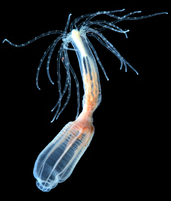
Researchers originally targeted Nematostella vectensis, the starlet sea anemone, in the search for an organism that could help them understand the evolution of complex bilaterian animals. Bilaterians, which include insects, worms and vertebrates, have body plans built on bilateral symmetry, with sensory organs at the anterior and centralized nervous systems. However, Nematostella is also capable of extensive regeneration, and the 2007 publication of its genome opened the door for the anemone to inform our understanding of animal evolution and regeneration.

“Some people describe sea anemones as these primitive animals,” says Michael Layden. “But you can use them to ask modern questions.”

Layden, an associate professor of biological sciences, uses Nematostella to try to answer questions related to nervous system evolution and to understand how neuronal regeneration differs from development, which has implications in how researchers think about regenerative therapy design.

“When I started [my career] … there were lots of good regeneration models,” he says. “Hydras had been around for almost 300 years, and planarians are these little crazy worms that you can cut into 211 pieces and get 211 worms. But the problem with those systems is that you cannot really get embryos, so you cannot study development effectively. The animals are incredible for learning about regeneration, but you cannot ask the specific question of how similar and dissimilar regeneration and development are. So that's what attracted me to Nematostella.”

Layden began his career studying nervous system development in Drosophila—the fruit fly, which he calls “the poster child for genetics”—at the University of Oregon. There, he encountered researchers interested in linking evolution and development—and discovered Nematostella, an organism more genetically similar to humans than Drosophila.

“I saw a talk on Nematostella, two in a row, actually, that came out of the lab that I ended up doing my postdoc in [at the University of Hawaii at Manoa],” he says. “What I realized was, first of all, this is a weird animal, but they're doing cutting-edge research on it. They are taking advantage of the new era in genomics and are able to ask pretty sophisticated questions. At that time, if it wasn't a mouse, a fly, a zebrafish or a frog, you couldn't do anything in it. But they were doing it in this sea anemone—they were knocking down genes and trying to look at function. And it just kind of hit me that I could still work on the nervous system, I could look at evolution and I could study regeneration, all in this one system.”

Layden, who first glimpsed research in action when he was a laboratory dishwasher as an undergraduate at the University of Rochester, has never looked back. He now helms the Layden Lab, which studies neural development in the starlet sea anemone.

“Even to this day, I'm still excited to come to work and see what we're going to learn,” he says.

Two Big Questions

Layden and his team aim to answer two questions: First, how was the ancestral nervous system that gave rise to the human brain patterned? And second, how can an organism remake neurons and rewire the nervous system during regeneration?

The answers to these questions may help in developing a better understanding of neurogenesis—the formation of neurons in the brain—and potentially aid in improved treatments of central nervous system disorders.

Image of the starlet sea anemone with black background

Nematostella vectensis, the starlet sea anemone, helps researchers understand animal evolution and regeneration.

To answer the first question, Layden and his team are trying to determine how different neurons first were made: “How did cells go from naive to neural? And then how do all the different types of neurons that an organism needs to sense its environment and determine the appropriate response get patterned?”

The mechanisms that patterned the ancestral nervous system can help researchers understand whether complex brains evolved once or multiple times, which has significant implications about our understanding of animal evolution, he explains.

“What we think is that by understanding how Nematostella patterns its nervous system [and how its relatives pattern theirs] … we can build a theoretical model of what the ancestral system looks like,” Layden explains. “That allows us to understand what might have occurred during nervous system evolution. What we're seeing is that patterning we thought was specific to complex brains is actually patterning Nematostella, too.”

Complex brains, including the human brain, are patterned from the anterior to posterior axis using a gradient of a Wingless-Int (WNT) protein. That gradient then makes the specific domains of the nervous system, which give rise to the forebrain, midbrain, hindbrain and spinal cord. That entire program is already present in Nematostella, says Layden.

“For basically 100 years or so, people have said that all brains look like they're patterned the same, so they evolved once. But really, of course they would look like they are patterned the same way, because they are just co-opting something that was already there. It wasn't specific to the brain. So that means that that information cannot be used to say that brains only evolved once.

“We're hoping that by looking at Nematostella, we can start to get a better sense of what we can look at in brain development that would be informative to tell us if evolution happened once or multiple times, and potentially how many times. … It's really simple at its core, but it took time for the technology of science to get to the point where people could think about it this way. We are part of that group of newer biologists trying to look at these older questions with new technology,” he explains.

Development & Regeneration

The second question—How do you remake the nervous system through regeneration?—requires a focus on how the neurons in a regenerated animal were made relative to the mechanisms that made those same neurons during embryonic development. Layden and his team use Nematostella to compare the processes of development and regeneration.

“It's becoming more apparent that during regeneration, the nervous system—or probably all tissues—is actually not being built with the exact same programs used to construct them during development,” Layden says. “It kind of makes sense because the way that animals are patterned is sort of context-specific, and regeneration is different from development. In hindsight it's obvious, but this is a question that researchers struggled with for a long time.”

This difference makes a comparison between development and regeneration even more necessary. “We need to know that difference because if you're trying to use regenerative therapies for biomedical intervention and you just force animals down this developmental pathway, it's sort of doomed to fail,” Layden explains. “And so, we are now rethinking our approach to regeneration research and regenerative medicine.”

During development and regeneration, each neuron undergoes a process that determines its job. This process requires turning parts of the organism’s genome on and off in specific ways, which is defined as a gene regulatory network, says Layden. Layden and his team build gene regulatory networks to determine the order that the genes in the Nematostella genome are turned on during development and regeneration of different types of neurons. They then compare the two processes.

Diffusion MRI of the human brain, purple and pink in color, black background

A diffusion MRI of the human brain. The Layden Lab is working to better understand neurogenesis—the formation of neurons in the brain.

The team has identified 20 neural genes—known as transcription factors—that act right after a cell switches from naive to neural to start each neuron down its own path during development. They then work to put these genes in order by reducing or taking away the function of one of them and observing what happens to the remaining genes. “So if you take something away and five of the other genes are now all of a sudden at higher levels, then you know that the function of the one you took away was to block the activity of those other five. And, conversely, if you take it away and you see these other 10 genes all are no longer expressed, then you know that the job of the particular gene you took away is to turn those other 10 genes on. Using that logic, we build a preliminary gene regulatory network describing the order in which the genes function,” says Layden.

Next, using biochemistry, molecular biology and genomics, the team tries to refine the regulatory networks that govern neuronal patterning.

Regeneration is more challenging to examine than development, says Layden.

“It's not like you have a separate subset of your genome that functions during regeneration and during development. They both are acting in both processes, but they are used in different ways. So if you get rid of a gene, development never happens and you can't study regeneration. Thus, to study regeneration, we need to design approaches that allow us to control whether a gene is disrupted during development or regeneration.

“The first thing we are doing is just making a map of where all our developmental genes are during regeneration. When we do that, we see some very simple patterns. Sometimes, where the gene is turned on during regeneration is different from where it's turned on during development. We know that it can't be functioning exactly the same, just based on that simple observation. We don't know how it's different yet, but we know that it's not the same.

“Other times it appears, based on the expression pattern, that a particular gene probably does something similar in both processes. So we are building that information first, and simultaneously trying to come up with new technologies to take away gene activity during regeneration. To do that, we are adapting tricks that have been used in traditional model systems,” he explains.

The complete answers to these big questions, Layden says, will be found beyond Nematostella.

“The answer isn't going to come from only using Nematostella. Development and regeneration should be compared in multiple species to identify the patterns in how these processes differ across the tree of life,” he says. “When we look broadly enough, patterns always emerge that shape our understanding of all biology and advance our ability to positively impact human health.”

Layden received a National Institutes of Health (NIH) R03 in 2016, an R01 award in 2019 and a National Science Foundation (NSF) CAREER Award in 2020 to support this work.

Credit Where It’s Due

When Michael Layden and postdoctoral research associates Layla Al-Shaer and Jamie Havrilak were preparing a book chapter about the history of Nematostella as a model system, Al-Shaer and Havrilak began a deep dive into the discovery of the sea anemone.

British naturalist and marine biologist Thomas A. Stephenson published the first description of Nematostella in 1935. Havrilak noticed that Stephenson attributed the anemone’s discovery to “Miss G.F. Selwood,” who first found the sea anemone at the Isle of Wight in England and sent the specimens to Stephenson.

“A lot of times in Nematostella papers you see Stephenson's name mentioned because he's the one that published the book describing them, but it's only if you dig through his really hard-to-find book that you realize that it was a woman who actually found them,” Havrilak explains. “And so we started this hunt for information about her.”

Al-Shaer searched ancestry websites for a photo of Selwood, and Havrilak reached out to an archivist at Selwood’s alma mater in the United Kingdom for more information. The archivist identified her as Gertrude Fanny Selwood, a lecturer in zoology at Municipal College who had received a bachelor’s degree at the University of Birmingham. Even with Selwood’s full name, however, Al-Shaer and Havrilak were unable to find any photos—but not for lack of trying.

“[When I lecture], I try to point out when a woman actually did the work and didn't get credit or if she worked for somebody and that person gets cited but she's the one who actually did [the work], just to highlight that women were doing the science,” says Havrilak. “Nematostella is becoming more established, but a lot of people have never heard of it or don't know a lot about it, so you give a brief history of the model when you're talking to a broad audience. Including Selwood in that history is important.”

Says Al-Shaer: “We really wanted to have a picture of [Selwood] to say, ‘Look, this woman was out there, trudging around in coastal estuaries, finding these species.’”

Story by

Kelly Hochbein

Related Stories

Anna Sullivan and Randi Conroy with PDI students on Field Day

Lehigh Students Win 2024 Davis Projects for Peace Award

The team aims to fight malnutrition in Bududa, Uganda, through a sustainable poultry program and education campaign.

small cities illustration

College of Arts and Sciences Initiative Aims to Help Small Cities

The college launches the Small Cities Lab to provide an interdisciplinary research approach to finding solutions to the problems facing small cities.

man intervenes in protest between two people from opposing sides

New Study on Psychology of Blame Points to Promising Strategies for Reducing Animosity in Political Divides

The study by Psychology Professor Michael Gill and Raihan Alam ’23 shows partisan animosity can be reduced by a historicist thinking intervention.