Elderly woman with glasses

Unpicking the Secrets of Neurodegenerative Diseases

Neuroscientist Daniel Babcock is using fruit fly research to pave the way to treatments for Parkinson’s and other diseases.

Story by

Mary Ellen Alu

Photography by

Christa Neu

Videography by

Stephanie Veto

Tags

Vials of fruit flies line the shelves of neuroscientist Daniel Babcock’s lab on the second floor of Iacocca Hall. Here, where fruit flies number into the tens of thousands, if not millions, Babcock and his students are creating models of neurodegenerative diseases such as Alzheimer’s and Parkinson’s in the flies to unpick the diseases’ secrets and potentially pave the way to treatments.

“One of the most common questions I get is, why would you look at fruit flies to study something as complex as neurodegenerative disease?” says Babcock, assistant professor of biological sciences.

But the tiny fruit flies—only a few millimeters long—remarkably share some 70 percent of the same genes as humans, which puts them in what Babcock considers a “sweet spot” of model organisms.

Fruit flies in a jar

Fruit flies share approximately 70 percent of the same genes as humans, making them model organisms for the study of disease.

“They are simple enough, where we can study thousands and thousands and thousands of fly brains,” he says. “But they are complex enough, where there is enough similarity between fly biology and human biology that if we model these human diseases…hopefully, whatever we find in how these diseases work in the flies will then somehow be relevant to human health and disease.”

For more than a century, the fruit fly—Drosophila melanogaster—has proven to be a tremendous resource in understanding many fundamental and important questions in human development. Six Nobel Prizes have been awarded for groundbreaking research using fruit flies, including the 2017 Nobel Prize in physiology or medicine that went to three scientists who used fruit flies in their discoveries about the molecular mechanisms controlling the body’s circadian rhythm.

“Our brains have somewhere around 100 billion neurons, the flies have about 250,000,” Babcock explains. “So that’s much less than ours, but it’s still a large group of neurons. Their neurons work the same way as ours do. They use the same chemicals to communicate with each other.”

Babcock and his team are focusing their research on three major areas of inquiry: Why are certain populations of neurons vulnerable to a particular disease? How do accumulations of protein aggregates spread throughout the brain? What is happening at the synapses of neurons in different neurodegenerative diseases?

In trying to answer the first question, Babcock and his team are focusing on why dopamine-producing neurons selectively die in patients with Parkinson’s disease, a progressive disorder of the central nervous system that affects movement. He says another set of neurons tends to be “ground zero” for Alzheimer’s, a progressive disorder that affects memory and other mental functions.

Different parts of the brain and different proteins are involved, and yet, time and time again, these different diseases all share this feature of aggregate accumulation.

Daniel Babcock

“So people who have a mutation that might render them vulnerable to Parkinson’s disease tend to lose dopaminergic neurons,” Babcock says. “But why are dopaminergic neurons the ones that are lost and not the ones that affect learning and memory, like it is with Alzheimer’s disease? ... We don’t have an answer for why that is.”

When the scientists manipulate the dopaminergic neurons in the flies’ brains or examine flies with mutations for PINK1 or Parkin—two genes closely linked to Parkinson’s—they find that the flies exhibit similar motor problems to humans, such as difficulty walking and climbing, Babcock says. By dissecting the flies’ brains and staining them with antibodies to make them “light up” in different colors, the scientists are able to analyze the flies’ brains under a microscope.

“One of the nice things about flies is they have such a short life span,” Babcock says. “They can live 60 to 70 days normally, but a 20- or 30-day-old fly is already pretty old,” so scientists don’t have to wait decades to be able to study the progression of a disease.

By the Numbers

60K

Americans diagnosed with Parkinson’s each year

1.5

The number of times men are more likely to have the disease than women

10%

Amount of those 65 and older living with Alzheimer’s

5.7M

Americans living with Alzheimer’s disease

6th

Leading cause of death in the United States

$1.1T

Projected cost to American society in terms of caring for those with Alzheimer’s, in 2018 dollars, by 2050

How Do Protein Aggregates Spread?

In another key area of research, Babcock and his team are looking at how protein aggregates spread throughout the brain.

Scientists have known for quite some time that neurodegenerative diseases are associated with aggregates forming in the affected neurons, he says. The protein alpha-synuclein can cause clumps known as Lewy bodies, the hallmark of Parkinson’s, for example. The plaques and tangles of Alzheimer’s that build up in the brain are accumulations of proteins called Beta-amyloid and Tau, respectively. Huntington’s and amyotrophic lateral sclerosis (ALS) are also associated with aggregates.

“Different parts of the brain and different proteins are involved, and yet, time and time again, these different diseases all share this feature of aggregate accumulation,” says Babcock. “We’ve thought for a while now that this clearly has some implication in disease progression.”

About a decade or so ago, when people started looking more carefully at where the aggregates started to accumulate, he says, they found that the aggregates seemed to follow neuronal circuits throughout the brain. That meant aggregates seen in one set of neurons would next be seen in a part of the brain that the neurons connected to.

“People thought, ‘Well, this seems to be a bit more than a coincidence. These aggregates are not just building up independently, they’re spreading throughout the brain, so they can actually travel from one cell to another,’” he says, “And that’s been a kind of scary prospect, that these aggregates can actually move and spread throughout the brain. We think that might be why, as these diseases progress, symptoms get worse and worse, because the pathology is literally spreading from one part of the brain to another.”

While at the University of Wisconsin, Babcock and genetics professor Barry Ganetzky used fruit flies to demonstrate that protein aggregates accumulated at synaptic terminals and progressively spread through the brain. Their study was published in 2015 in the PNAS (Proceedings of the National Academy of Sciences).

At Lehigh, Babcock and his team are further investigating how those aggregates are getting out of those neurons, and also, just as importantly, how they’re getting into other neurons. They are working on a model of Huntington’s disease, but they are building flies in which they can investigate other disease models, such as Parkinson’s, Alzheimer’s and ALS. Do those aggregates spread the same way as the Huntingtin aggregates do?

“It’s my favorite kind of experiment,” Babcock says, “because no matter what happens, it’s interesting. If they all end up spreading the same way, then anything we find with our Huntingtin aggregates could now be applicable to all these other diseases as well. If they don’t all spread the same way, that’s interesting for its own reason. Ok, why do these aggregates behave this way and these do not?”

What Happens at the Synapses?

In a third area of inquiry, Babcock and his team are trying to understand what happens at the synapses of neurons in neurodegenerative diseases. That’s important, he says, because of evidence that synaptic dysfunction and other problems at the distal ends of cells begin long before the neurons actually die.

“We think that losing neurons is actually a very late stage of the disease progression,” Babcock says. “It’s possible that by the time you’re actually losing neurons, it might be too late to really fix anything.”

With currently no cures for any of the major neurodegenerative diseases, Babcock is testing the hypothesis that what is taking place at the tips of the neurons is setting off a cascade of events that ultimately cause the neurons to die. “If we can understand what takes place much earlier, can we intervene at that point?” he asks. “Can we fix it and stop the neurons from dying?”

Important Research for an Aging Population

Babcock’s work in fruit fly research began “almost on a whim” while choosing rotations as part of his graduate study. He had already done several rotations with different researchers to get a better feel for their work—“because it’s a major commitment, spending five, six, seven years of your life” in a particular field. He decided on a final rotation with Michael Galko, of The University of Texas, MD Anderson Cancer Center, who used fruit flies to conduct pain research.

Fluorescent image of a fruit fly brain

Fruit flies share approximately 70 percent of the same genes as humans, making them model organisms for the study of disease.

“And I absolutely fell in love with it, stuck with it, continued to do that,” Babcock says. His post-doctoral work in fruit fly research into neurodegenerative diseases was at the University of Wisconsin. At Lehigh, his active research team includes graduate and undergraduate students and a post-doctoral fellow.

Hopefully, he says, the team will be able to find “something new” in how the proteins that are known to be involved with [neurodegenerative diseases] actually work, in trying to figure out a way to stop cell death, and specifically, how aggregates move from cell to cell throughout the brain, “which is fascinating and terrifying at the same time, that these toxic proteins can actually travel throughout the nervous system and spread throughout the brain,” he says.

“If we could find a condition either using a genetic manipulation or some type of tool where we can figure out how that spreading is happening mechanistically and then find a way to stop it, could we potentially identify some potential therapeutic target or drug target?” he asks. That, he says, would allow researchers who design drugs to treat patients “to have something they could try and aim for, instead of just designing compounds in the dark.”

Babcock notes that with people living longer, the potential of more and more people developing neurodegenerative diseases looms larger.

“This is not only a fascinating area of research,” he says of his team’s work, “but something where we can hopefully make a real difference that will matter to people.”

The Hows of Fruit Fly Research

by: Stephen Gross

The objective is clear: Daniel Babcock's students typically set out to model different types of neurodegenerative diseases in fruit flies. But how exactly do students go from creating the flies they want to study to actually getting them under the microscope?

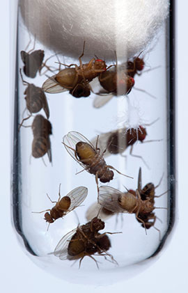
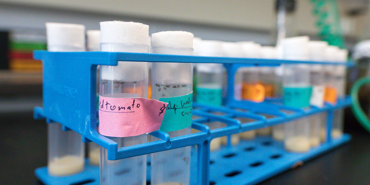
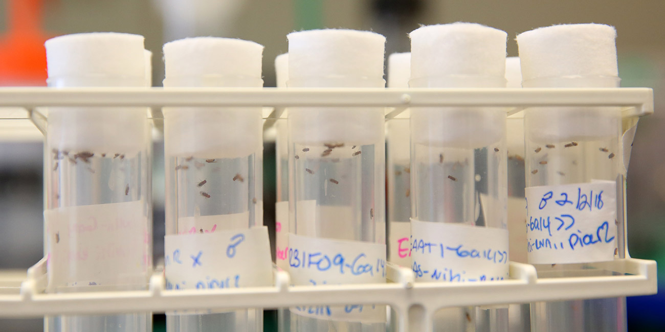
Knowing the age of the flies is crucial, so the first step is seeding a new vial. Researchers use CO2 to anesthetize several flies from the breeding stock, place them on a pad, and add to a new vial a number of males and females.

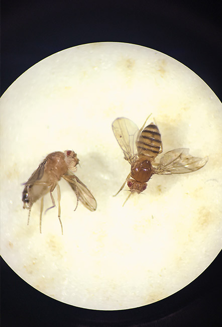
Once embryos develop, it takes about 10 days for adult flies to emerge, which are then collected and momentarily anesthetized with CO2 and sorted by gender.

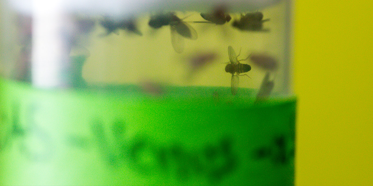
"They can get a little bit scrappy when you have males and females together," Babcock says. "They will fight over resources, they will fight over mates."

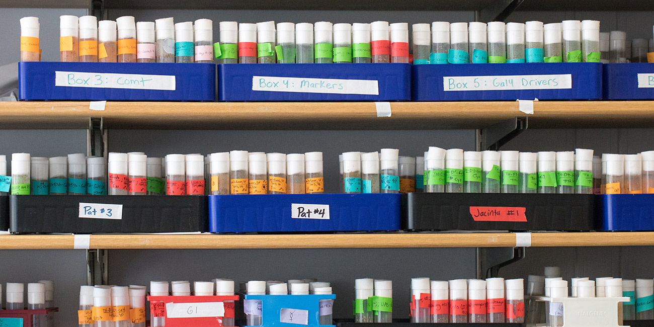
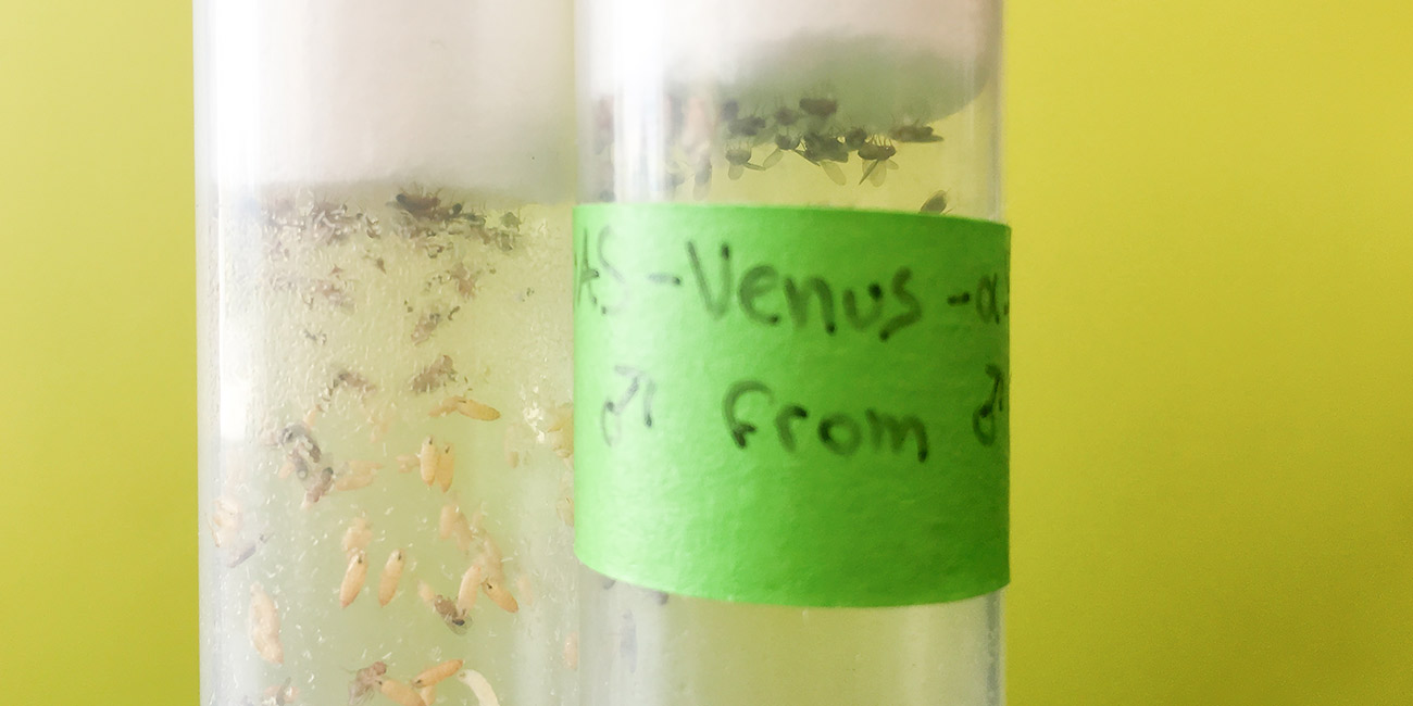
The flies are then aged, and researchers change the vials every few days to keep their food fresh.

Once the flies reach 20 or 30 days old, some will begin to show symptoms of a disease mutation, like Parkinson's. Other "wild-type" flies are used as a control. Researchers then dissect the fly brains they want to examine.

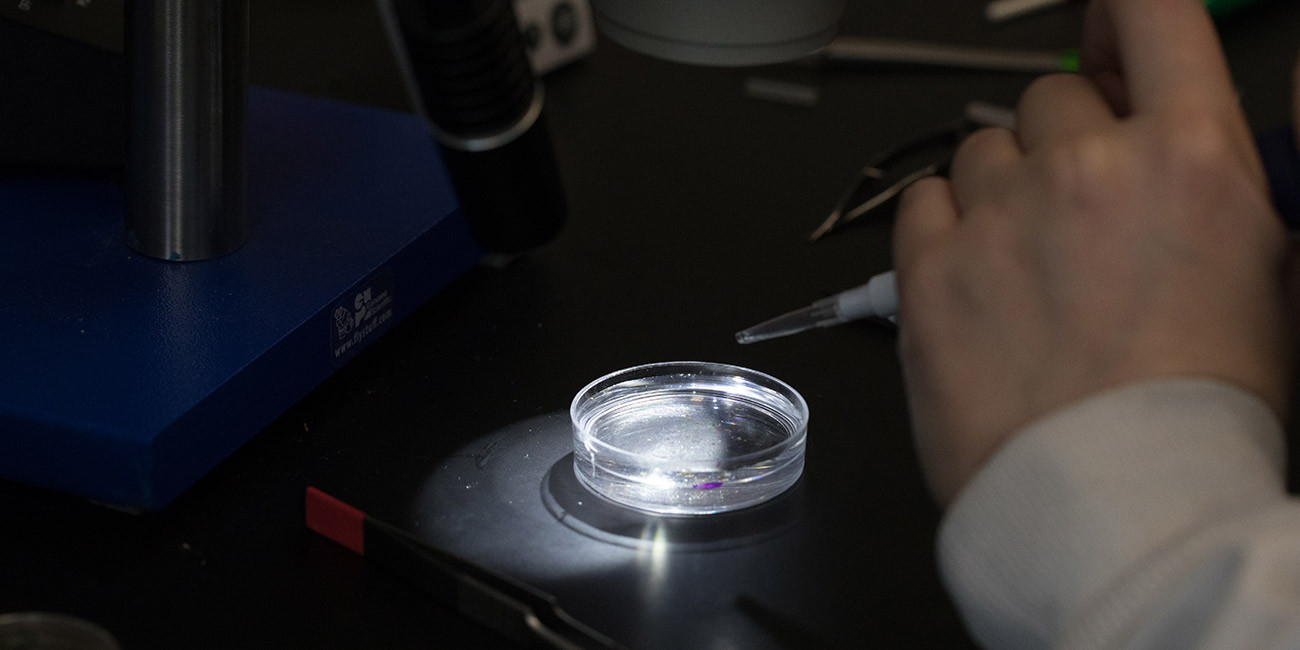
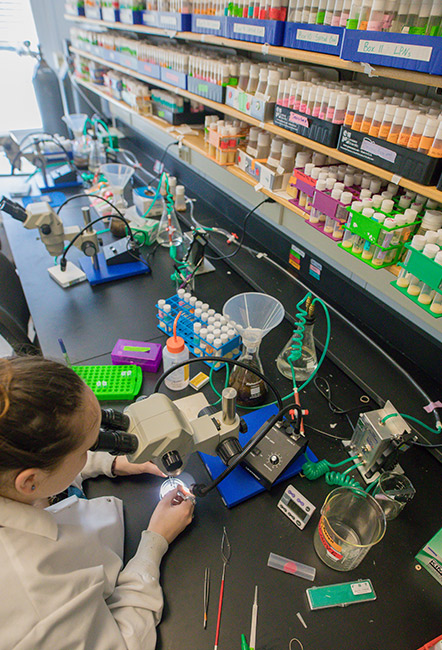
The selected flies are once again anesthetized with CO2 and transferred to a petri dish with a saline solution called PBS. The solution keeps the tissues from deteriorating and maintains a stable environment until the brains can be preserved. Brain dissection, through which 10 to 20 brain samples per condition are collected, occurs under a microscope.

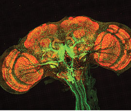
Before a researcher can examine the brain, they must preserve it with formaldehyde. Once preserved, the brain can be labeled with different antibodies. These antibodies can then be tagged with fluorophores of different colors, allowing particular proteins to light up within the brain.

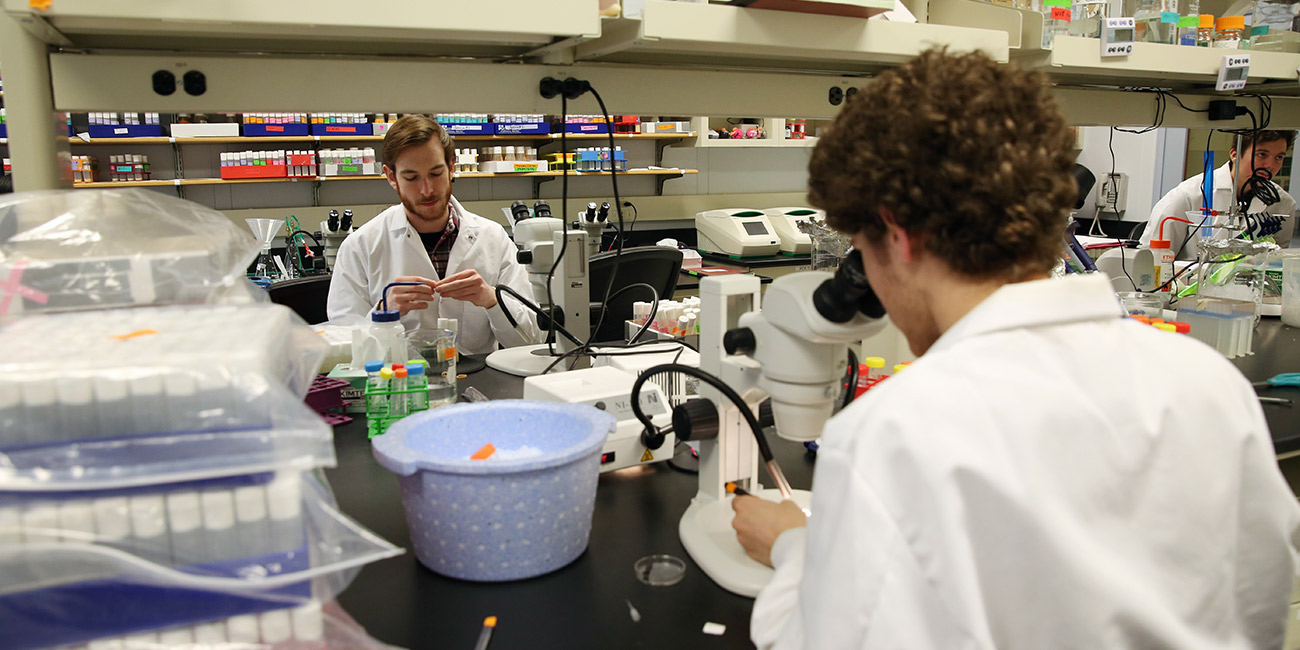
Finally, researchers wash off the brain samples, place them on a microscope slide and cap them with a coverslip. Then, fluorescent images can be taken under the microscope.

Daniel Babcock earned his Ph.D. in neuroscience from the University of Texas Health Science Center at Houston/MD Anderson Cancer Center. He did his post-doctoral work at the University of Wisconsin- Madison. The Babcock Lab at Lehigh is interested in understanding the cellular and molecular mechanisms underlying the earliest hallmarks of neurodegenerative diseases.

Story by

Mary Ellen Alu

Photography by

Christa Neu

Videography by

Stephanie Veto

Tags

Related Stories

flags behind a fence

Study: Threat of Deportation Leads to Psychological Distress Among Both Latino Citizens and Noncitizens

Amy Johnson and research collaborators find it’s not just undocumented immigrants who feel at risk.

Nicole Johnson research

New Research Highlights Link Between School Shootings and Violence Against Women

Research led by Nicole Johnson, associate professor of counseling psychology, finds that 70% of school shooters have perpetrated violence against women and can influence prevention strategies.

Santiago Herrera and a student at the symposium

Lehigh Oceans Research Center Holds Inaugural Symposium

The College of Arts and Sciences launches a new research center that focuses on the field of ocean science.