Better Detection of a Deadly Parasite
The life of a watershed is complex. The watershed is the area of land separating the smaller water flows that feed into a larger, common outlet—like a river, lake or ocean. Watersheds are often home to a variety of wildlife—and subject to agricultural and recreational uses as well. From this complicated ecosystem, bacteria, viruses and parasites emerge and sometimes make their way into the water supply.
Drinking water and public swimming pools are treated with chlorine is to kill such disease-causing organisms. Chlorine successfully eliminates a number of harmful pathogens, but not all of them.
Cryptosporidium parvum is one such chlorine-resistant pathogen. A protozoan parasite that is transmitted through direct contact or ingestion of contaminated food or water, C. parvum is found in animal and human fecal matter.
According to the Centers for Disease Control and Prevention, C. parvum is a leading cause of waterborne disease among humans in the United States. Infection can lead to cryptosporidiosis which, in healthy people, causes severe diarrheal disease that can last for weeks. For the elderly, for infants or for the immunocompromised, the infection can be deadly.
Because contaminated drinking water has been linked to major outbreaks of cryptosporidiosis, detecting and removing C. parvum in source waters is vital to protecting public health.
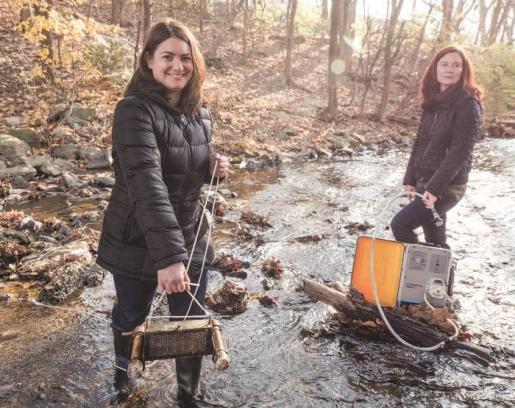
Two Lehigh engineers are investigating how C. parvum oocysts— the development stage in which the parasite exists in the environment—attach to environmental biofilms, which are the microbial communities that grow on rocks and other underwater surfaces. Their goal is to develop an improved detection method that can identify a contamination source faster and more cheaply than current methods, preventing widespread infection.
“We think we can develop a surface that can be commercially produced for around $5,” says Kristen L. Jellison, associate professor of civil and environmental engineering. “This would allow utilities and water resource managers to better protect the public from exposure by sampling more locations with greater frequency and obtaining more reliable and comprehensive data about the sources of the parasite in the watershed.”
Jellison and Sabrina S. Jedlicka, associate professor of materials science and engineering, are the first to demonstrate that the attachment of oocysts to environmental biofilms is a calcium-mediated process—a crucial step toward their goal. They describe their results in an article in Applied and Environmental Microbiology titled “Pseudo-second-order calcium-mediated Cryptosporidium parvum oocyst attachment to environmental biofilms.”
Understanding the mechanism that enables oocysts to attach to biofilms is important for calculating the attachment efficiency, or the rate at which the parasite binds to the material. Understanding the rate is key for two reasons. First, it may enable calculation of oocyst concentration in the water based on numbers of oocysts attached to a material. Second, it is needed to design materials that maximize oocyst attachment. Such attachment-enabling material will make it possible to detect the source of the infestation faster and more inexpensively than current methods allow.
“The number of oocysts on a given biofilm sample reveal how much of the parasite is present in the water,” says Jellison. “For example—simply for illustration’s sake – say that five oocysts attach to a biofilm downstream of the contamination source. That may indicate the presence of 100 oocysts further upstream. If we see ten oocysts attaching, it might mean there are 200 at the main source.”
Jellison and Jedlicka’s study provides new insights into the impact of calcium on the attachment of C. parvum oocysts to environmental biofilms. In addition, their modeling method could be used to elucidate the behavior of oocysts in common complex aquatic systems—an important step in enabling future innovations in parasite detection and treatment technologies to protect public health.
Comparable data, much cheaper
The idea for using environmental biofilms to identify a C. parvum contamination source first occurred to Jellison during her work with the Philadelphia Water Department on the detection of Cryptosporidium in the Wissahickon and Schuykill watersheds.
The EPA-approved filtration-based methods used to test the water sources were expensive and the results were inconsistent. One filter, says Jellison, could cost as much as $120. That may not sound like a large sum, but considering the limited resources of many municipal water departments—and the number of filters that might be needed to accurately detect the source of a C. parvum contamination—it represents a serious obstacle.
“Recovery using current methods depends on variables such as the cleanliness of water and the skill of the person doing the testing,” says Jellison. “C. parvum can be present in the water in very small amounts. You can check a 10-liter sample and not detect the parasite. But it could be present in the water.”
While testing watersheds near Philadelphia, Jellison scraped biofilms off rocks upstream and downstream of a suspected C. parvum contamination location and then tested the samples in her lab at Lehigh. Over time, the biofilm samples provided comparable data to the approved filtration method—but at much lower cost.
“Being able to test the water accurately and in more places increases the likelihood of identifying the contamination source and decreases the chance that thousands of people will become infected,” she says.
Jellison’s downstream biofilm samples contained more oocysts than the samples taken from rocks upstream of suspected point sources. However, there was no way to identify when the oocysts attached. The clues provided by the biofilms were important for confirming that C. parvum was entering the water at particular locations, but not much help in revealing when the contamination events occurred.
Jellison reasoned that if she could figure out precisely when the oocysts attached, she would be better able to identify patterns of oocyst contamination which would enable more effective source water protection strategies.
Designing materials to maximize attachment
That is where Jedlicka comes in. Jedlicka—a materials scientist who says she “comes from a field of designing surfaces to elicit behavior in cells”—does mathematical modeling that enables the duo to calculate the attachment efficiency of C. parvum to biofilms.
“We need an understanding of the chemistry behind the binding process to know what designs will work best,” says Jedlicka.
Jedlicka’s ability to calculate the binding force is what led to the demonstration of a calcium-mediated binding process.
“When we took the calcium out, the binding efficiency went down significantly, which is one way we knew that calcium is important to binding,” she says.
In addition to contributing to improved detection methods, creating a surface design that maximizes the attachment of oocysts has other implications as well.
“If we can identify the binding mechanism that enables C. parvum to ‘stick’ to biofilms, we can create an improved means to remove it,” says Jellison. “Understanding the mechanism could even open the door to preventing the parasite from binding in the intestine and making people sick.”
Among the team’s next steps is investigating the other chemical processes involved in the binding, in addition to calcium. They also plan to test their model under a number of conditions, including hydrodynamic sheer stresses.
Their ultimate goal, however, remains clear.
“If we can help utilities find a way to monitor multiple points in the watershed instead of just a few, they can put their limited resources to more effective use,” says Jellison.
“Until then,” says Jedlicka, “we’ll just keep asking ridiculously hard science questions.”
The research is supported by grants from the National Science Foundation, and by the Pennsylvania Department of Community and Economic Development through the Pennsylvania Infrastructure Technology Alliance.
Story by Lori Friedman
Photo by Ryan Hulvat
Posted on:


