

Mathematics: Modeling Aneurysms
Cerebral aneurysms (CA) lead to strokes for more than 40,000 Americans annually. Currently, there exists no formal framework toward the diagnosis of CAs, and most go unnoticed until they rupture or are detected by brain imaging that may have been obtained for another condition. Additionally, assessing the risk of a potential rupture of a diagnosed CA is mostly experiential as the biomechanics governing the time evolution of CAs is not fully understood. Developing a mathematical model to better understand the processes leading to and during a CA is the focus of research by Yue Yu.
Yu, assistant professor of mathematics, constructs arterial geometry from patient-specific CT data to understand the flow patterns that occur inside an aneurysm that has formed on the wall of one of the arteries. Visualization of the blood flow can reveal the complex flow patterns in the aneurysm, while rendering the stress tensor on the arterial walls highlights the local stresses in the aneurysm due to the flow structure interactions.
“The numerical simulation is challenging because the patient-specific aneurysm geometry is very complicated, which requires a high resolution in space. Therefore, the numerical solver has to be both high-performance and high-fidelity,” says Yu. “Also, the relatively light density of the arterial wall brings a further challenge: the convergence of the partitioned fluid (blood)-structure (arterial wall) interaction solver is problematic because of the so-called added-mass effect, and a new numerical algorithm needs to be designed.”
The challenge for researchers is there are few in vivo measurements with which they can compare and validate software. Current technologies only allow researchers to study aneurysms at specific moments in time. Yu hopes to develop software that will allow scientists to simulate aneurysm processes over longer periods of time.
Yu’s work is shedding new light on the conditions for arterial rupture. Developing a better model will allow researchers to zoom in and excerpt high-resolution images of the areas affected by blood flow and derived quantities that may be of vital importance to physicians. With improved images, they can clearly identify and treat the regions of high stress.
Posted on:
Thursday, April 09, 2015
Share This Story:


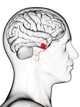
Modeling of cerebral aneurysms is providing new information about circumstances surrounding arterial rupture.